![Transcription factories in a Hela cell [from Cook PR (1999) Science 284, 1790]](../images/pombo.png)

('15mtmebm'; Peter.Cook@path.ox.ac.uk; http://users.path.ox.ac.uk/~pcook)
Objectives
The material covered in this lecture, coupled
with the recommended reading, should enable you to:
• understand the concept of a cell cycle
• describe the processes of mitosis and meiosis,
and the essential differences between them
• understand that recombination allows genes
to be shuffled, so new combinations can be tested by evolution.
Mitotic cycle
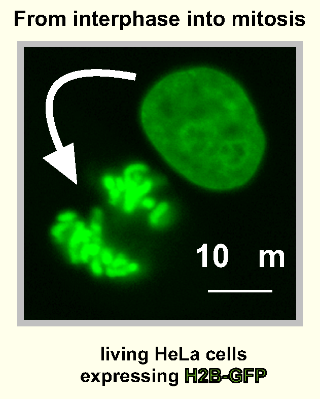
During interphase, chromatin forms dense mass, but during cell division
(mitosis) condenses into discrete chromosomes (Fig).
Mitosis divides parental cell equally between 2 daughters; mitochondria and
vesicles derived from ER segregate randomly, chromosomes
precisely.
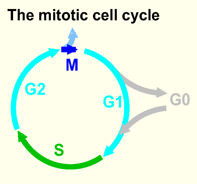
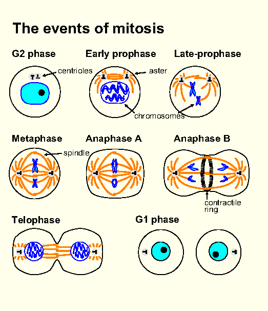
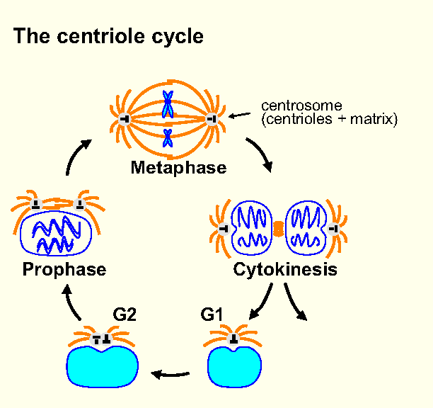
Cell cycle (Fig): G1, S (Fig), G2, M (Fig). G0 (non-dividing, differentiated, cells); programmed cell death, apoptosis;
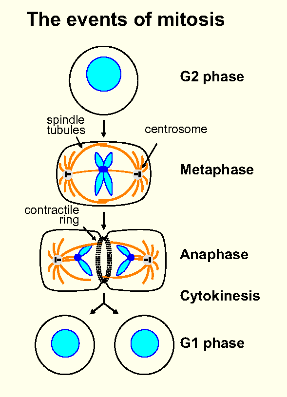
S originally recognized using [3H]thymidine; M visible in LM (nuclear membrane breaks down, chromatin condenses into chromosomes,
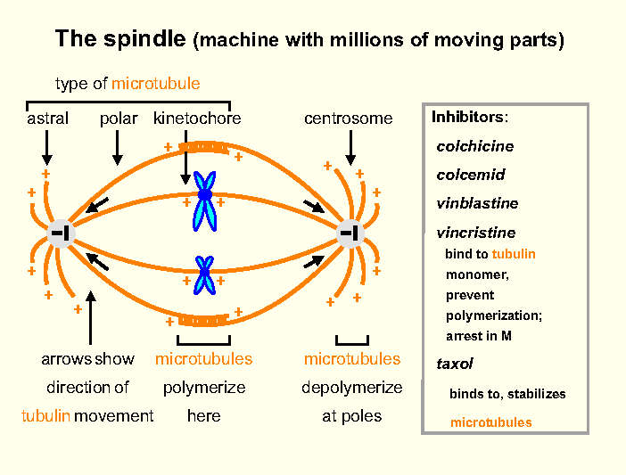
attach to spindle containing tubulin (Fig), segregated accurately
by spindle, contractile ring (actin) pinches cell
in two; Fig). Spindle inhibitors (Fig).
M divided: pro-, meta-, ana- (A and B), telo-phase (Fig).
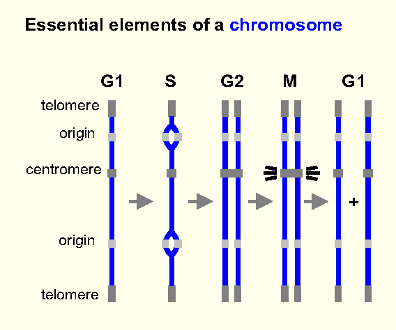
Metaphase chromosome: 2 chromatids, centromere (spindle, correct segregation), telomere (Fig).
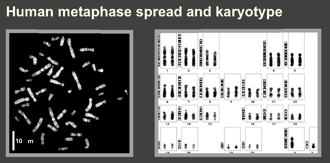
Classify: metacentric (centromere in middle), acrocentric (towards
one end), telocentric (at end). Alternatively: sex + autosomes (44 autosomes + 2 X in female, or 1 X + 1 Y in male). Karyotype (46 in human, 40 in mouse; Fig).
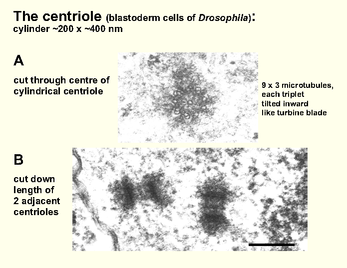
Centrosome - densely-staining pair of centrioles surrounded
by amorphous matrix (Fig); radiating microtubules (microtubule organizing center, aster); split and moves during cycle (Fig); some tubules catch centromeres, then spindle pulls apart the 2 chromosome
sets.
Meiotic cycle (Fig)
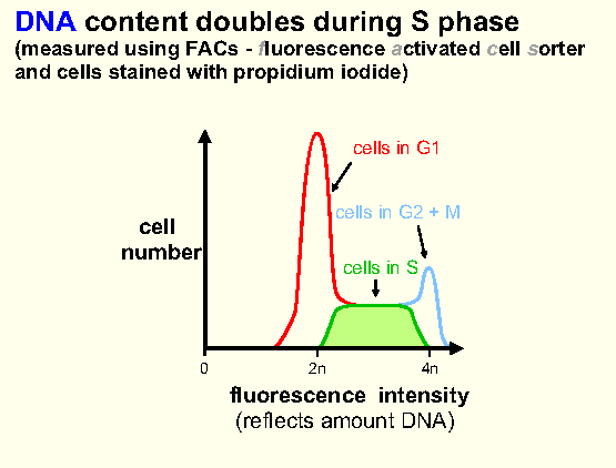
Somatic cell - diploid (2n) amount of DNA in G1, tetraploid (4n) in G2. Sexual reproduction: production of haploid (1n) gametes
during meiosis, fusion of 2 gametes to give (2n) zygote.
DNA of 2n cell duplicated, then 2 successive nuclear divisions generate 4
haploid gametes. Fertilization restores diploidy. So alternation of haploid
+ diploid generations. In most higher organisms, haploid phase brief.
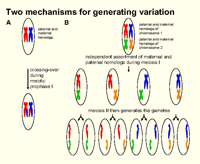
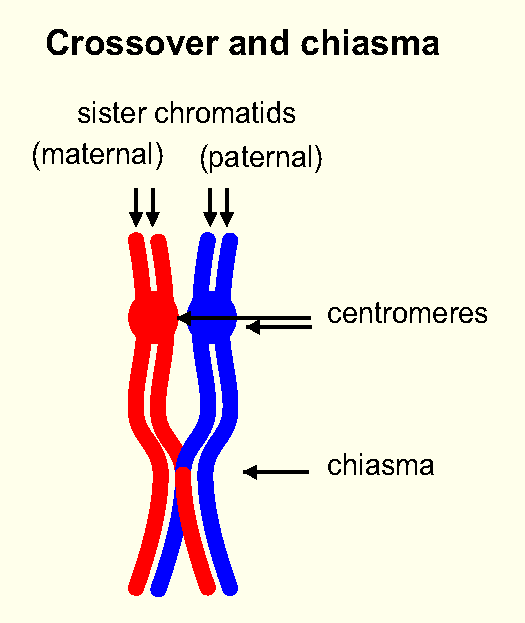
Two features meiosis ensure haploid generation receives mixed
set genes (increased genetic variation): (i) segments homologs exchanged (recombined) randomly, (ii) maternal/paternal
homologs assorted (segregated) semi-randomly at 1st division (Fig).
New gene combinations tested by evolution - fit survive/reproduce, less fit culled.
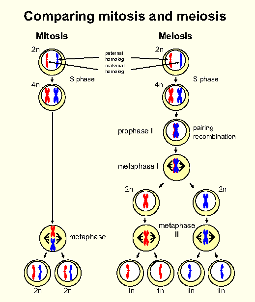
Meiosis as 3-stage process: DNA of 2n cell replicated to
generate chromosomes with attached chromatids in 4n cell, chromosomes divided
among 4 haploid cells by consecutive divisions (meiosis I, II). During
I (reductional division), homologs pair, segregate (pairing
required for proper segregation). During II (equational division), sisters segregated.
Almost invariably, each of resulting 4 haploid cells differs genetically from
other 3. Variability arises because (i) new variants generated by recombination, (ii) chromosomal
set split semi-randomly among haploid cells, and (iii) additional variation
introduced during fertilization when different male/female gametes fuse; therefore,
each diploid egg carries unique set genes. First source of variation arises
from high levels of homologous recombination that occur during
I. Bivalent, join (chiasma, chiasmata; Fig).
Second source discovered by Mendel - semi-random segregation that occurs during I (segregation not random in that each haploid
cell eventually receives only 1 copy of each homolog, but completely random
in that different homologs segregate independently; Fig).
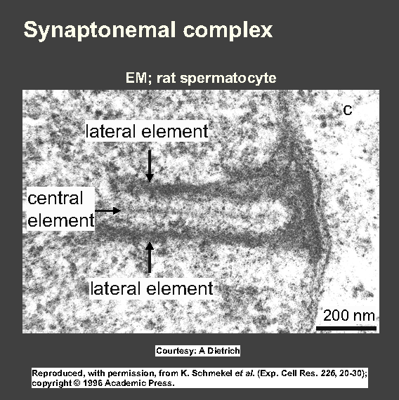
Stages of I. Prophase (can occupy 90% meiosis) when duplicated
chromosomes partially condense to become visible as chromosomes, dance, 'feel' among crowd for partners. Once partners identified
(leptotene), homologs pair or synapse (zygotene), tightly align (synaptonemal complex during pachytene; Fig), unpair (desynapse during diplotene) and
move apart (diakinesis; Fig). When I ends, cells in many organisms
pass quickly through II. No DNA made during this interval, and
II similar to mitosis, except only 1 (duplicated) copy of
each chromosome present.
Recombination
Important roles in most cells (eg in bacteria, main role enables replicating
complex to bypass lesion in parental strand by exchanging daughter templates),
but central role in meiosis (rate >1000x that in mitotic cycle).
Meiotic recombination: DNA sequence exchanged between homologs, shuffling of different versions of genes (alleles)
so new combinations tested by evolution. Different organisms
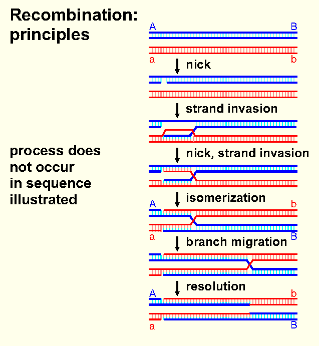
use different pathways, share principles (Fig):
(i) 2 homologous DNA duplexes cross-over (double helices break, broken
ends join). (ii) Initiation: homology search (DNA-DNA pairing).
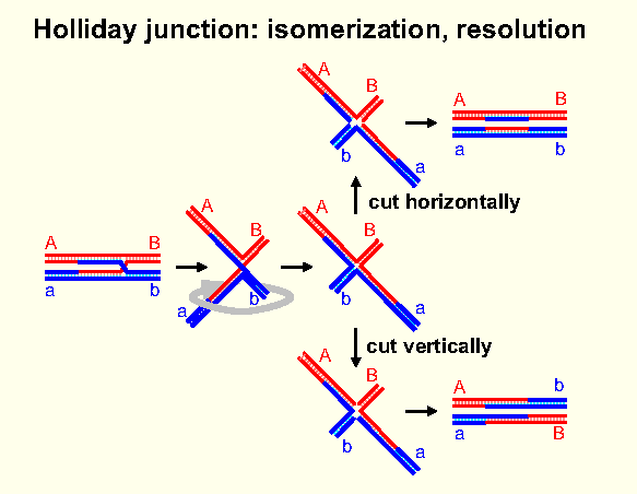
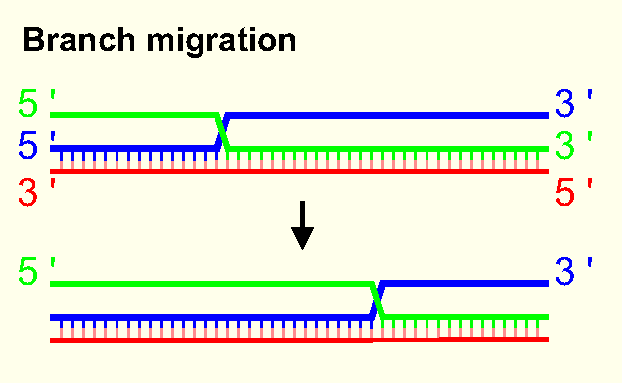
(iii) Gives staggered heteroduplex joint (strand in one duplex paired
with complementary strand in other) - key X-shaped Holliday
junction (Fig), branch migration (Fig). (iv) Cleavage/rejoining generally precise, so sequence
at point of exchange remains unchanged (reciprocal v non-reciprocal
recombination). (iv) Overall effect is to move genes between homologs, without
normally changing relative positions on chromosome.
References
Ch 17, 21 in Alberts, B. et al. (2014). 'Molecular
Biology of the Cell'. 6th Ed. Garland [see also PubMed].
Hochwagen, A. (2008). Meiosis. Curr. Biol. 18, R641-645. [PubMed]